


With osteochondrosis of the spinal column, many are known not by the gears known from the TV screen, but from their sad experience.Statistics are harsh: up to 80% of the population suffers from this disease, which also significantly younger.If previous complaints about back problems were mainly among the older generation, now no one is more surprising.And the fault of a sedentary lifestyle and the so -called "benefits of civilization".

The cervical spine osteochondrosis is polyetiological a progressive disease that manifests with degeneration of intervertebral discs and the dystrophy of the ligamentous apparatus of the spine.Everyone knows about first -hand symptoms, but these knowledge is fragmentary;We will try to structure them as well as talk about the principles of diagnosing and treating osteochondrosis of the cervical back.
Causes of osteochondrosis
Medical science cannot respond unequivocally, which is why osteochondrosis occurs.It is certain that the sedentary lifestyle that a modern person is inclined to adversely affect the progression of this disease.It is interesting that both the hypodynamia and colossal loads of athletes lead to disk representatives.An inheritance factor plays a key role.The following reasons are distinguished:
- Inheritance story charged;
- Obesity;
- Hippodinami;
- metabolic disorders in the body;
- traumatic damage to the spinal column;
- long static overloads and lifting weights (computer work, weight lifting, miners, moving, etc.);
- scoliosis;
- dysfunctional environmental situations;
- flat feet and pregnancy;
- Hypothermia and stress, which often cause irritation of the disease.
There are several neurological syndromes:
- Shoulder periartrit;
- root;
- cardiac;
- Vail artery syndrome.
Shoulder periartrit.It is characterized by pain in the neck, shoulder, shoulder joints.The main neurogenic contraction of the shoulder joint is formed, which is natural protective, as it protects the axillary nerve from extension (antalgic pose).With this position, the muscles surrounding the joint are in tension.The severity of the pain syndrome depends on the degree of deterioration of osteochondrosis: a small restriction of amplitude of movements in the joints in the so -called "frozen shoulder" when any movement causes severe pain.The pain intensifies when the shoulder is diverted and pronounced, as it is these movements that increase the pressure of the axillary nerve.
Royshift syndrome (cervical radical).Most often it occurs with cervical osteochondrosis.At the same time, the spine of the spinal nerve is squeezed due to the "decrease" of the intervertebral discs, as well as the increase in osteophytes or the extension of the discs in the lateral direction.Pain syndrome is specific: intense burning, tearing, pressing pain, which also intensifies when the patient moves the head.Antalgic pose is also observed in the neck muscles, they are severely tense and painful, the volume of movements is limited.There is pain in the back of the head, neck, front chest, shoulder, between shoulder blades.The composition of sensitivity from the type of "half of short -sleeved jackets" is characteristic.
Cardial syndrome.The name of the syndrome is responsible for himself: clinical photography is very similar to pectoris angina.In this case, there is no organic heart damage, at the peak of pain syndrome, coronary blood flow violations by ECG are not detected, and such patients are well tolerated.A typical feature with angina pectoris: the pain is done after taking nitrates, and in the case of osteochondrosis does not change and disturb for a long time.Unlike angina pectoris, the localization of pain is mainly in the heart to the left.With the irritation of the roots of the C8 - T1 segments, rhythm concerns in the form of tachycardia and extrasitol are possible.This is not due to damage to the heart system, but with a violation of sympathetic intrigue of the heart muscle (extracardiac damage).In the differential diagnosis of angina pectoris and cardiac syndrome, leadership is the fact that, in addition to cardiac complaints, the patient notes the increase in pain in the shoulder and neck fusion associated with the rise or severe movements.
Vail artery syndrome.The vertebral artery is done in a channel formed by holes in the transverse processes of the vertebrae.This artery is paired, it is responsible for the blood supply of the brain.Therefore, any narrowing of this channel has a very negative impact on brain tissue nutrition.Vertebral artery syndrome develops directly with both the compression of the artery itself and with the irritation of the sympathetic nerve plexus, which is around it.Pain in this pathology is burning or pulsating in the occipital region with spread in whiskey, tutorial arches, crops.It arises from one and both sides.Patients usually associate deterioration with post -sleeping states of non -physiological position, transport trips, walking.With pronounced symptoms, hearing loss, dizziness, ears noise, nausea, vomiting, loss of consciousness and increased blood pressure are possible.Such symptoms are not specific and are very similar to complaints in the cerebral stroke.This pathology is characterized by the Sistine hat syndrome: a pallor that occurs when you crash your head back (severe brain ischemia).He was described by visitors to the Sistine Chapel to the Vatican when they examined the frescoes in its arches.It is also possible to fall without loss of consciousness with sharp head curves.
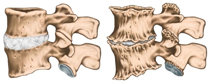
Like any diagnosis in medicine, the diagnosis of osteochondrosis is established on the basis of complaints of patients, anamnesis of the disease, clinical examination and auxiliary research methods.X -cervical spine in direct and lateral projections is performed, if necessary in special positions (with an open mouth).At the same time, experts are interested in the height of the intervertebral discs, the presence of osteophytes.For modern research methods, IAMR and CT research is used, which make it possible to verify the diagnosis more accurately.In addition to the listed methods of additional research, consultation of related specialists (cardiologist, ophthalmologist, neurosurgeon) may be necessary, and the neurologist examination is purely vital.The neurologist is engaged in the treatment of osteochondrosis, so after examining the patient, he will prescribe the minimum examination needed at his discretion.
The treatment of osteochondrosis
Osteochondrosis is a polyetological disease, for a course of therapy does not cure.You cannot drink a "magic pill" and everything will pass, it is necessary to radically change the lifestyle of your life, as the trigger is the hypodynamia.The most vulnerable results are easier to reach at the initial stage of the disease, when complaints are minimal and there are no compression syndromes and spinal arteries.In the acute phase of the disease, when the following groups of medication are described by pronounced pain: Pain syndrome is pronounced:
- Preverstebral therapeutic blockade (to relieve pain and removal of muscle spasm);
- NSAIDS;
- ointments containing NSAID and reflex action;
- muscle relaxants;
- B Vitamin V.
As the inflammatory process softens and relieves pain syndrome, they move on to the treatment of physiocherapy.Most often, the following techniques are used:
- Laser therapy;
- electrophoresis;
- acupuncture;
- Exercise therapy;
- therapeutic massage;
- Manual therapy.
It is important to understand that osteochondrosis continues with periods of irritation and forgiveness, so it is very important to influence, and not to treat the investigation.